
Fig. 1
- ID
- ZDB-FIG-231215-128
- Publication
- Touret et al., 2022 - A dual involvement of Protocadherin-18a in stromal cell development guides the formation of a functional hematopoietic niche
- Other Figures
- All Figure Page
- Back to All Figure Page
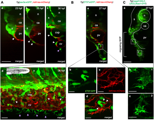
Stromal-endothelial cell interactions during SCP migration and caudal vein plexus formation. (A-C) Confocal spinning-disk images of live embryos of the indicated transgenic backgrounds and developmental stages. (Aa-Ac) Optical transverse sections reconstituted by Imaris software. White arrows indicate SCPs migrating on the primordial vein or within the forming CVP; purple arrow indicates an FMC. Scale bar: 30 µm. (Ad) Lateral view. Schematic indicates the region shown. GFPhigh and GFPdim cells are neural crest-derived pigment cells and SCP derivatives, respectively. Scale bar: 20 µm. (Ba) Optical transverse section. (Bb-Bd) Magnified projections from three confocal planes spaced by 0.6 µm at the region indicated by the dashed square in (Ba), ventral to the primordial vein, where angiogenesis is building the CVP. White arrows point to stromal cell interaction with endothelial cells via filopodia. Scale bars: 10 µm. (Ca) Optical transverse section. SCPs first migrate in a 2D mode in close contact with the primordial vein (dashed square marked 1) then in a more 3D mode when they co-migrate with endothelial cells (dashed square marked 2). Scale bar: 20 µm. (Cb,Cc) Magnified images of SCPs in 2D (Cb) and 3D (Cc) migration modes. Scale bars: 10 µm. ca, caudal artery; cv, caudal vein; cvp, caudal vein plexus; n, notochord; pv, primordial vein; s, somites. |