FIGURE

Fig 6

ID
ZDB-FIG-231213-23
Publication
Forti et al., 2023 - Identification and impact on Pseudomonas aeruginosa virulence of mutations conferring resistance to a phage cocktail for phage therapy
Other Figures
All Figure Page
Back to All Figure Page
Fig 6

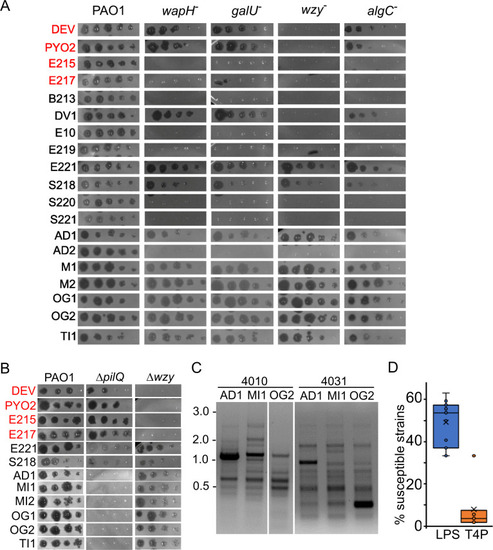
Phage growth on different P. aeruginosa strains. (A and B) Plating on PAO1 mutants. Phage lysates were diluted (×10) in 96-well plates and replicated on PAO1 and the indicated PAO1 mutants. The plates were incubated overnight at 37°C. wapH, PAER5b; galU, PAER6b; wzy, PADR6; algC, PAER10b, ΔpilQ, PAMO302; Δwzy, PAMO301. The CK4 components are reported in red. (C) Different RAPD PCR band pattern obtained with primers 4010 and 4031 on the indicated phages. The same patterns were obtained in a replicate experiment. MW marker migration is indicated on the left (kb). (D) Percentage of the P. aeruginosa strains listed in Table S4 susceptible to all phages listed in Table 4 but E221, for which only a subset of clinical strains was analyzed. Each dot represents the host range of a different phage. LPS, phages not growing on wzy mutants; T4P, phages not growing on the ΔpilQ mutant.

Expression Data

Expression Detail
Antibody Labeling
Phenotype Data

Phenotype Detail
Acknowledgments
This image is the copyrighted work of the attributed author or publisher, and ZFIN has permission only to display this image to its users. Additional permissions should be obtained from the applicable author or publisher of the image. Full text @ Microbiol Spectr