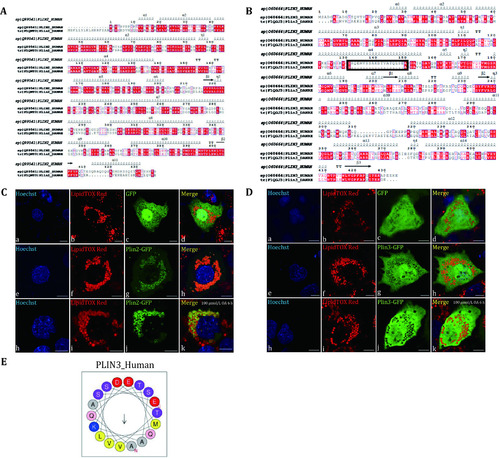
Localization of Plin2 and Plin3 of zebrafish in Huh7 cells. A The amino acid sequence alignment of Plin2 of human and zebrafish. The amino acid sequences were compared by Clustal X and visualized by ESPript3.0. B The amino acid sequence alignment of Plin3 of human and zebrafish. The amino acid sequences were compared by Clustal X and visualized by ESPript3.0. The black frame showed the absence of sequence in zebrafish Plin3. C Zebrafish plin2 was cloned into GFP plasmid and transiently overexpressed in Huh7 cells. The cells were imaged by an Olympus FV1200 confocal microscope. Upper panels: the vector GFP overexpressed in Huh7 cells; Middle panels: Plin2-GFP overexpressed in Huh7 cells; Lower panels: Plin2-GFP overexpressed in Huh7 cells and treated with 100 μmol/L OA for 6 h. Green: GFP; Red: LipidTOX Red; Blue: Hoechst. Bar = 10 μm. D Zebrafish plin3 was cloned into GFP plasmid and transiently overexpressed in Huh7 cells. The cells were imaged by an Olympus FV1200 confocal microscope. Upper panels: the vector GFP overexpressed in Huh7 cells; Middle panels: Plin3-GFP overexpressed in Huh7 cells; Lower panels: Plin3-GFP overexpressed in Huh7 cells and treated with 100 μmol/L OA for 6 h. Green: GFP; Red: LipidTOX Red; Blue: Hoechst. Bar = 10 μm. E Helical wheel projection of sequences of human PLIN3 in the black frame was predicted by HeliQuest
|
