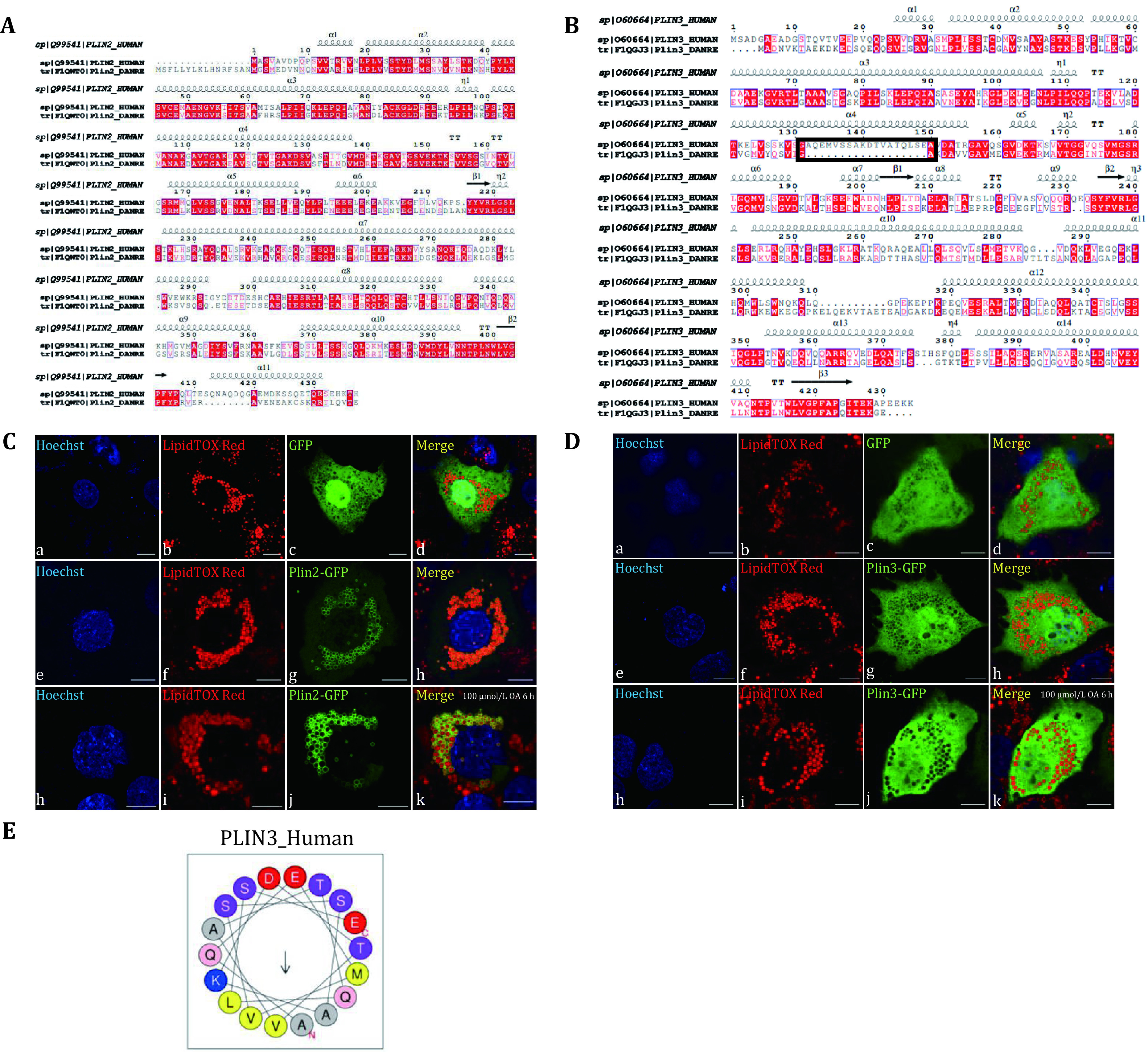
Localization of Plin2 and Plin3 of zebrafish in Huh7 cells. A The amino acid sequence alignment of Plin2 of human and zebrafish. The amino acid sequences were compared by Clustal X and visualized by ESPript3.0. B The amino acid sequence alignment of Plin3 of human and zebrafish. The amino acid sequences were compared by Clustal X and visualized by ESPript3.0. The black frame showed the absence of sequence in zebrafish Plin3. C Zebrafish plin2 was cloned into GFP plasmid and transiently overexpressed in Huh7 cells. The cells were imaged by an Olympus FV1200 confocal microscope. Upper panels: the vector GFP overexpressed in Huh7 cells; Middle panels: Plin2-GFP overexpressed in Huh7 cells; Lower panels: Plin2-GFP overexpressed in Huh7 cells and treated with 100 μmol/L OA for 6 h. Green: GFP; Red: LipidTOX Red; Blue: Hoechst. Bar = 10 μm. D Zebrafish plin3 was cloned into GFP plasmid and transiently overexpressed in Huh7 cells. The cells were imaged by an Olympus FV1200 confocal microscope. Upper panels: the vector GFP overexpressed in Huh7 cells; Middle panels: Plin3-GFP overexpressed in Huh7 cells; Lower panels: Plin3-GFP overexpressed in Huh7 cells and treated with 100 μmol/L OA for 6 h. Green: GFP; Red: LipidTOX Red; Blue: Hoechst. Bar = 10 μm. E Helical wheel projection of sequences of human PLIN3 in the black frame was predicted by HeliQuest
Acknowledgments
This image is the copyrighted work of the attributed author or publisher, and
ZFIN has permission only to display this image to its users.
Additional permissions should be obtained from the applicable author or publisher of the image.
Full text @ Biophys Rep
Your Input Welcome
Thank you for submitting comments. Your input has been emailed to ZFIN curators who may contact you if
additional information is required.
Oops. Something went wrong. Please try again later.