|
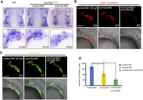
cxcl12a mutants exhibit reduced pharyngeal pouch cells. (A) Expression levels of nkx2.3 and cv2 expression were assayed in wild-type or cxcl12a mutants post-injection with either control MO or cxcl12b MO. (B–C) Confocal microscopy elucidates images showing pharyngeal pouch cellular architecture in wild-type and cxcl12a, or combined cxcl12a and cxcl12b knockdown embryos within a Tg(nkx2.3:mCherry)(B) or Tg(sox17:GFP)(C) background at 36 hpf. Scale bar, 50 μm. (D) Quantification of mCherry-positive cells within the Tg(nkx2.3-mCherry) transgenic line was executed from a sample size of nine embryos. Error bars are indicative of standard deviation (SD). Statistical significance denoted by *, p < 0.05; **, p < 0.01, as gauged by Student’s t-test.
|