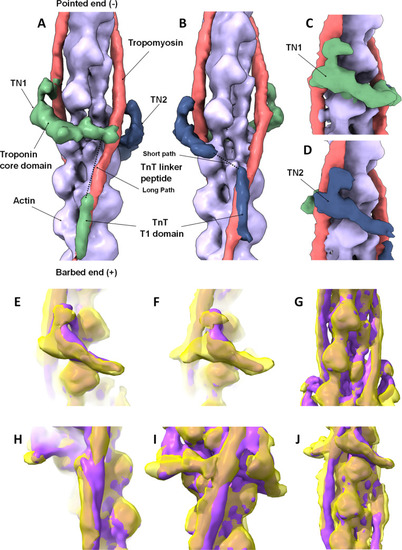
3D reconstruction of native zebrafish cardiac thin filaments and comparison to human reconstituted thin filament structure. A-D: Surface rendered protein density map, highlighting the thin filament constituent proteins, segmented and colour coded as follows; Tn1 green, Tn2 blue, tropomyosin pink and actin purple. A & B: views of the complete map oriented to illustrate the two distinct paths taken by individual troponin molecules on each side of the thin filament as they span the two tropomyosin strands. A & B are related by 180o rotation about the central axis of the thin filament. The different paths of troponin are apparent; Tn1 is located higher on the filament than Tn2, however, the TnT linker peptide path of Tn1 to tropomyosin is longer than that of Tn2. More density is recovered for Tn1 despite its linker peptide following the longer path. C & D: Close up views of the troponin core domain illustrating its rotated ‘L’ shape which is consistent on both sides. Views as in A & B but rotated by 60o about the central axis. Segmented regions calculated using ChimeraX. The pointed (-) and barbed end (+) of the actin filament are indicated. E–J: The zebrafish (yellow) and Yamada (purple) high Ca2+ state reconstructions are superposed. E & F: Extra protein density in Tn1 and Tn2 core regions is visible in the zebrafish map. G & J: Tropomyosin and actin densities are similar. H: Strong similarity in TnT T1 domains interacting with tropomyosin overlap region. I: Extra TnT linker-region density in the zebrafish map not present in other thin filament maps
|
