
FIGURE
Fig. 4
- ID
- ZDB-FIG-230919-11
- Publication
- Ruggiero et al., 2023 - FSCN1 as a new druggable target in adrenocortical carcinoma
- Other Figures
- All Figure Page
- Back to All Figure Page
Fig. 4
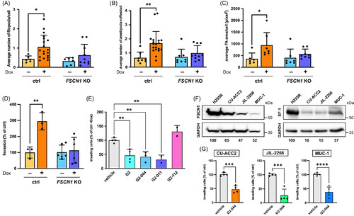
FSCN1 inactivation or pharmacological inhibition reduces in vitro Matrigel invasion of ACC cells. Number of (A) filopodia, (B) lamellipodia/ruffles and (C) area of focal adhesions per cell in control and FSCN1 KO H295R cells treated with either vehicle or Dox (1 μg/ml) for 24 h. n (independent experiments) = 6-9. Mean ± SD is shown. *P < .05; **P < .01, one-way ANOVA with Šidák's multiple comparisons test. (D) In vitro Matrigel invasion of control and FSCN1 KO H295R cells treated with either vehicle or Dox (1 μg/ml). Results are expressed as percentages of control cell invasion. n (independent experiments) = 3-6. Mean ± SD is shown. **P < .01, one-way ANOVA with Šidák's multiple comparisons test. (E) In vitro Matrigel invasion of control H295R cells treated with Dox (1 μg/ml) and with vehicle or G2 (50 μM), G2-044, G2-011 or the inactive G2-112 compound (all 5 μM). n (independent experiments) = 2-4. Mean ± SD is shown. **P < .01, one-way ANOVA with Šidák's multiple comparisons test. (F) Left: immunoblot showing expression of FSCN1 and GAPDH in H295R, CU-ACC2, JIL-2266 and MUC-1 cells. FSCN1 levels (relative to GAPDH) in the CU-ACC2, JIL-2266 and MUC-1 ACC cell lines are indicated as percentages of H295R, which express the highest levels of FSCN1. Right: immunoblot showing expression of SF-1 and GAPDH in H295R, CU-ACC2, JIL-2266 and MUC-1 cells. SF-1 levels (relative to GAPDH) in the CU-ACC2, JIL-2266 and MUC-1 ACC cell lines are indicated as percentages of H295R, which express the highest levels of SF-1. FSCN1 and SF-1 band intensities were quantified by Image J after GAPDH normalization. n (independent experiments) = 3. G) Matrigel invasion of those ACC cell lines treated with vehicle or G2-044 (5 μM). n (independent experiments) = 4-5. Mean ± SD is shown. ***P < .001; ****P < .0001, t test.
|
Expression Data
Expression Detail
Antibody Labeling
Phenotype Data
Phenotype Detail
Acknowledgments
This image is the copyrighted work of the attributed author or publisher, and
ZFIN has permission only to display this image to its users.
Additional permissions should be obtained from the applicable author or publisher of the image.
Full text @ Int. J. Cancer