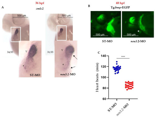
Downregulation of neu3.2 affects cardiac development. (A) Analysis of the cardiac marker cmlc2 by whole-mount in situ hybridization (WISH) in zebrafish embryos injected with STD-MO and injected with neu3.2-MO (1 pmol/embryo). Images are the anterior views of the whole mount with heart with dorsal up. Arrows point to the atrium (a) and ventricle (V), demonstrating their small size compared with the STD-MO-injected embryos. Ratios at the bottom left part of each picture specify the number of embryos showing the same staining pattern, compared to the total number of embryos used for each experiment. Two replicates were performed (n = 35). (B) Representative images of the cardiac area in the Tg(Bmp:EGFP) line STD-MO-injected and neu3.2-MO-injected (1 pmol/embryo at 48 hpf) embryos. Asterisk points out the reduction of fluorescence intensity and size of the heart in both conditions. Results are from one representative experiment with at least 25 embryos out of three independent replicates. (C) The graph shows the comparison of the heart rate (numbers of beats per minute) between STD-MO-injected and neu3.2-MO-injected (1 pmol/embryo) embryos and it is representative of one experiment (n = 25). The experiment was repeated three times. Results are expressed as the mean ± SD of three independent experiments. (**** p < 0.001 vs. the control group—embryos injected with standard morpholino-STD-MO).
|
