
Figure 3
- ID
- ZDB-FIG-230916-204
- Publication
- Zilliox et al., 2023 - Protocol to locally express cxcl12a during zebrafish olfactory organ development by combining IR-LEGO with live imaging
- Other Figures
- All Figure Page
- Back to All Figure Page
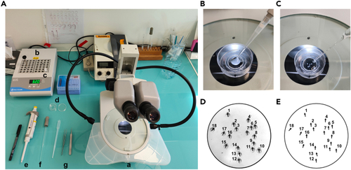
Mounting the zebrafish embryos in 0.7% low melting agarose (A) Material used for mounting embryos. (a) Stereoscopic microscope equipped with a transmitted light source. (b) Heating block set at 32°C. (c) Small aliquot of 0.7% LM agarose in fish water. (d) Small Petri dish. (e) 200 μL pipette. (f) Fine tip glass Pasteur pipet. (g) Dissecting tweezers. (B) Pipet 120 μL of heated 0.7% LM agarose in fish water and drop it in the center of the small Petri dish. (C) Transfer the dechorionated embryos to the agarose drop using a glass Pasteur pipet. (D) Picture showing the orientation of the embryos (black arrowhead points towards the anterior part, the head, of the embryo). (E) Embryos positions are annotated on the lid. |