FIGURE

Fig. 4

ID
ZDB-FIG-230814-121
Publication
Tang et al., 2021 - The H3K27 demethylase controls the lateral line embryogenesis of zebrafish
Other Figures
All Figure Page
Back to All Figure Page
Fig. 4

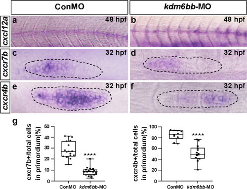
The chemokine signaling pathway is disrupted by kdm6bb-MO. a–f Representative in situ staining of chemokine members cxcl12a, cxcr4b, and cxcr7b are compared between the controls and kdm6bb morphants at 32–48 hpf. (a, b)The consecutive expression of cxcl12a is interrupted in Kdm6b knockdown embryos, both (c, d) cxcr4b and (e, f) cxcr7b expressions are downregulated in kdm6bb morpholino mutants in comparison with the control-MO-injected embryos. g The ratio of cxcr7b and cxcr4b-positive cells to total cells in the primordia of controls (ConMO; n = 18 embryos for cxcr7b, and n = 10 embryos for cxcr4b) and experiment group (kdm6bb-MO; n = 19 embryos for cxcr7b, and n = 14 embryos for cxcr4b). The black dotted lines outline the primordium appearance. The leading region is positioned to the right and the trailing region is to the left. Data are recorded as mean (minimum and maximum values). ****p < 0.0001

Expression Data

Expression Detail
Antibody Labeling
Phenotype Data

Phenotype Detail
Acknowledgments
This image is the copyrighted work of the attributed author or publisher, and ZFIN has permission only to display this image to its users. Additional permissions should be obtained from the applicable author or publisher of the image. Full text @ Cell Biol. Toxicol.