
FIGURE
Fig. 1
- ID
- ZDB-FIG-230803-21
- Publication
- Owen et al., 2023 - Loss of the crumbs cell polarity complex disrupts epigenetic transcriptional control and cell cycle progression in the developing retina
- Other Figures
- All Figure Page
- Back to All Figure Page
Fig. 1
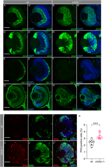
The effect of crb2a−/− on retinal neurogenesis and lamination. Characterisation of cell types present in zebrafish retina at 56 hpf (A–H, wild type zebrafish, A′-H′ crb2am289 zebrafish). Early retinal progenitor cells were identified through staining with pax6 in WT (A and B) and crb2a−/− (A′ and B′) and anti-vsx2 (C, D, C′ and D′) at 56 hpf. Anti-ZPR1 antibody (zpr1) was used to identify the presence of cone cells (E and F) in WT; however these were absent from the mutant retina (E′ and F′) at 80 hpf. The presence of Müller cells was visualised using an anti-glutamate synthetase antibody (GS) in WT (G and H) and mutant (G′ and H′) retina. M-phase nuclei, visualised using an anti-phospho-Histone 3 antibody (PH3), were observed in both WT (I) and crb2a−/− (J) at 56-hpf. All nuclei are stained with DAPI. S-phase nuclei as visualised by 5-ethynyl-2’-deoxyuridine (EdU) incorporation were observed in WT (I′) and crb2a−/− (J′) retina. Panels I″ and J″ are merged images of previous panels. (K) Quantification of PH3-positive cells; *** p < 0.001. Scale bar, 50 μm.
|
Expression Data
| Antibodies: | |
|---|---|
| Fish: | |
| Anatomical Terms: | |
| Stage: | Long-pec |
Expression Detail
Antibody Labeling
Phenotype Data
| Fish: | |
|---|---|
| Observed In: | |
| Stage: | Long-pec |
Phenotype Detail
Acknowledgments
This image is the copyrighted work of the attributed author or publisher, and
ZFIN has permission only to display this image to its users.
Additional permissions should be obtained from the applicable author or publisher of the image.
Full text @ J. Pathol.