FIGURE
Fig. 8
- ID
- ZDB-FIG-230719-35
- Publication
- Salehin et al., 2022 - Ventricular Anisotropic Deformation and Contractile Function of the Developing Heart of Zebrafish in-vivo
- Other Figures
- All Figure Page
- Back to All Figure Page
Fig. 8
|
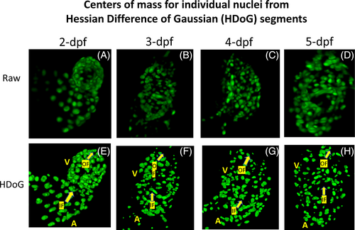
Visualizing spatiotemporal dynamics of myocardial cardiomyocyte nuclei from zebrafish at 2, 3, 4, 5 dpf. (A, B, C, D) Volumetric reconstruction of myocardial cardiomyocyte architecture acquired using high magnification water objective lens, enabling higher spatial resolution albeit with tradeoff in field of view. (E, F, G, H) Segmented nuclei volumes using Hessian Difference of Gaussian (HDoG) filter in conjunction with watershed algorithm. A, atrium, IF: inflow through AV valve, OF: outflow through VB valve; V, ventricle. Arrow heads point to the direction of the flow |
Expression Data
Expression Detail
Antibody Labeling
Phenotype Data
Phenotype Detail
Acknowledgments
This image is the copyrighted work of the attributed author or publisher, and
ZFIN has permission only to display this image to its users.
Additional permissions should be obtained from the applicable author or publisher of the image.
Full text @ Dev. Dyn.

