
Fig. 1
- ID
- ZDB-FIG-230718-1
- Publication
- Berg et al., 2022 - Brainstem circuits encoding start, speed, and duration of swimming in adult zebrafish
- Other Figures
- All Figure Page
- Back to All Figure Page
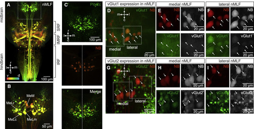
Separate molecular and anatomical nMLF neuronal populations (A) Orthogonal projection of confocal images showing reticulospinal neurons retrogradely labeled by biotinylated dextran injection in the spinal cord (IRF, inferior reticular formation; IMRF, intermediate reticular formation; SRF, superior reticular formation). Dorso-ventral position is color coded (v, ventral in red and d, dorsal in blue). (B) Enlarged image of the nMLF (dashed box in A) showing four cells individually identified across animals (MeM, MeLm, MeLr, and MeLc). (C) Top panel shows that nMLF neurons express the transcription factor Pitx2c. Middle panel shows neurobiotin (NB) retrogradely labeled nMLF neurons from the spinal cord (red). Bottom panel is the merge of Pitx2c expression and neurobiotin showing double labeled nMLF neurons. (D) Orthogonal projection of confocal images showing the differential expression of vGlut1 in nMLF neurons. (E) Lack of expression of vGlut1 in medial nMLF neurons indicated by the dashed box “medial” in (D) (arrows indicate the position of neurobiotin [NB] labeled neurons). (F) Expression of vGlut1 in lateral nMLF neurons indicated by the dashed box “lateral” in (D) (arrows indicate the position of neurobiotin [NB] labeled neurons). (G) Orthogonal projection of confocal images showing the differential expression of vGlut2 in nMLF neurons. (H) Strong expression of vGlut2 in medial nMLF neurons indicated by the dashed box “medial” in (G) (arrows indicate the position of neurobiotin [NB] labeled neurons). (I) Weak expression of vGlut2 in lateral nMLF neurons indicated by the dashed box “lateral” in (G) (arrows indicate the position of neurobiotin [NB] labeled neurons). |