FIGURE

Fig. 1

ID
ZDB-FIG-230625-29
Publication
Samimi et al., 2023 - Light-sheet autofluorescence lifetime imaging with a single-photon avalanche diode array
Other Figures
All Figure Page
Back to All Figure Page
Fig. 1

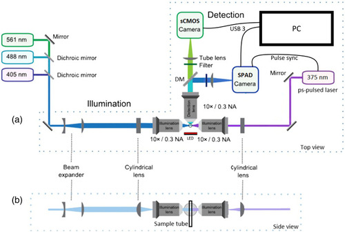
Schematic of the light-sheet FLIM microscope (adapted with permission from Ref. 52). (a) Top view of the SPIM system. The right illumination arm uses a 375 nm ps-pulsed diode laser operated at 50 MHz repetition rate to excite NAD(P)H fluorescence via a 200  μm wide light sheet, while the time-resolved SPAD camera images the NAD(P)H autofluorescence lifetime (after a 495 nm LP DM and a 440/80  nm filter) and the sCMOS camera images the NAD(P)H intensity (using a 530/55  nm filter). The left illumination arm uses a bank of CW lasers to excite other fluorophores via a 1.5 mm wide light sheet, while emissions are imaged on the sCMOS camera. A red LED provides trans-illumination for bright field imaging. (b) Side view of the beams in the illumination arms and the orientation of the sample tube. DM, dichroic mirror.

Expression Data

Expression Detail
Antibody Labeling
Phenotype Data

Phenotype Detail
Acknowledgments
This image is the copyrighted work of the attributed author or publisher, and ZFIN has permission only to display this image to its users. Additional permissions should be obtained from the applicable author or publisher of the image. Full text @ J. Biomed. Opt.