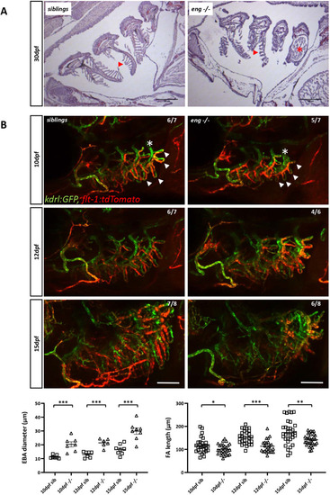
Abnormal branchial vascular development in Endoglin-deficient fish underlies structural impairment of gills. (A) H&E-stained histological sections of 30 dpf sibling and eng−/− gills. Note the poorly developed lamellae (arrowheads) and enlarged artery (asterisk) on branchial arch 4 (AA6) in eng−/− fish gills. Scale bars: 100 µm. (B) Top three rows: kinetic analysis of gill vascular development in 10, 12 and 15 dpf sibling and eng−/− fish in Tg(kdrl:GFP), Tg(flt1:tdtomato) background. Note the reduced length of afferent filamental artery (GFP+, Tomatohigh) and efferent filamental artery (GFP+, Tomatolow/−) (arrowheads) and enlarged efferent branchial artery) (asterisks) as early as 10 dpf. Note the overall loss of flt1 signal in 15 dpf eng−/− fish. Scale bars: 100 µm. Bottom row: graphical representation of sibling and eng−/− fish branchial efferent artery (EFA) diameter (left) and filamental artery (FA) length (right) at 10, 12 and 15 dpf. EFA diameter and FA length were measured on AA6. EFA diameter was measured at the most dorsal FA level. FAs (five per fish) were measured starting from the most dorsal FA. Data are presented as individual values and mean±s.e.m. EFA diameter: siblings (sib) versus −/−, ***P=0.0003, ***P=0.0006 and ***P=0.0003 at 10, 12 and 15 dpf, respectively. FA length: sib versus −/−, *P=0.037, ***P<0.0001 and **P=0.0026 at 10, 12 and 15 dpf, respectively. One-tailed Mann–Whitney test.
|
