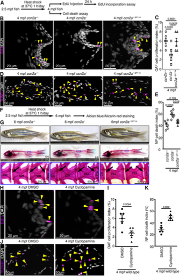
Ectopic expression of a constitutively active form of Fgfr1 can restore cellular phenotype in adult ccn2a−/− IVDs through SHH signaling. (A) Schematic depiction of experimental procedures. (B) Maximum intensity projections (MIPs) of confocal images of sagittal IVD sections stained for EdU (magenta; proliferating cells) and nuclei (white). Arrowheads indicate EdU+ cells in the OAF. (C) Quantification of proliferating OAF cell (n=6). (D) MIPs of confocal images of sagittal IVD sections stained for TUNEL (magenta; dead cells) and nuclei (white). Arrowheads indicate TUNEL+ cells in the NP. (E) Quantification of NP cell death (n=6). (F) Schematic depiction of experimental procedures. (G) Bright-field lateral views of live and AB/AR stained zebrafish. Black arrows indicate intervertebral spaces. Scale is marked in millimeter intervals. (H) MIPs of confocal images of sagittal IVD sections stained for EdU (magenta; proliferating cells) and stained with DAPI (white; nuclei). Arrowheads indicate EdU+ cells in the OAF. (I) Quantification of proliferating OAF cells (n=6). (J) MIPs of confocal images of sagittal IVD sections stained for TUNEL (magenta; dead cells) and nuclei (white). Arrowheads indicate TUNEL+ cells in the NP. (K) Quantification of NP cell death (n=6). In C,E,I,K, data are mean±s.e.m.; each sample represents one animal. Digits on the images in G indicate the number of fish that showed the presented phenotype out of the total number of fish. ‘C’ on images in G represents centrum/vertebrae.
|
