
FIGURE
Fig. 2
- ID
- ZDB-FIG-230522-11
- Publication
- Shin et al., 2023 - A heterozygous mutation in UBE2H in a patient with developmental delay leads to an aberrant brain development in zebrafish
- Other Figures
- All Figure Page
- Back to All Figure Page
Fig. 2
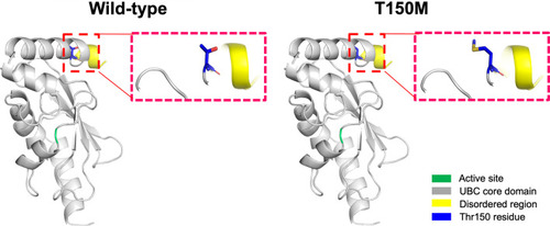
Structure homology-modeling of the normal human UBE2H and T150M variant. By means of in silico protein structure modeling, wild-type and mutant residues (p.Thr150Met) in the UBE2H protein have been represented as sticks alongside the surrounding residues. The Thr150 residue is located in the UBC core domain away from the active site. The crystal structure of the domain from wild-type UBE2H was generated using SWISS-MODEL (https://swissmodel.expasy.org/) and has been depicted as a cartoon representation |
Expression Data
Expression Detail
Antibody Labeling
Phenotype Data
Phenotype Detail
Acknowledgments
This image is the copyrighted work of the attributed author or publisher, and
ZFIN has permission only to display this image to its users.
Additional permissions should be obtained from the applicable author or publisher of the image.
Full text @ Hum. Genomics