
Fig. 3
- ID
- ZDB-FIG-230515-105
- Publication
- Potts et al., 2022 - Splicing factor deficits render hematopoietic stem and progenitor cells sensitive to STAT3 inhibition
- Other Figures
- All Figure Page
- Back to All Figure Page
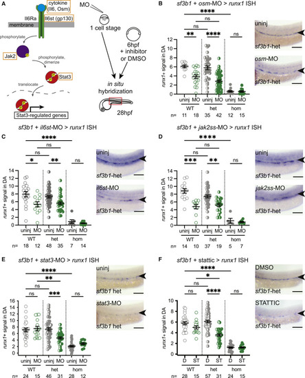
Zebrafish Sf3b1 heterozygous mutant HSPCs are sensitive to STAT3 inhibition (A) Schematic of Jak/Stat signaling pathway (left). Genes targeted for knockdown are indicated (orange box). Experimental strategy (right): target genes were knocked down in zebrafish embryos derived from sf3b1 heterozygote incrosses via either morpholino (MO) injection (one-cell stage) or small-molecule inhibitor administration (6–28 hpf), then samples were fixed at 28 hpf for in situ hybridization with HSPC marker runx1. (B–F) Representative in situ hybridization images of runx1 signal in dorsal aorta (DA) (arrowheads) of sf3b1 heterozygous (het) embryos (right) and quantification of DA runx1 signal in embryos with dots representing individual embryos (left). Targets tested include: ligand osm via morpholino (B); co-receptor il6st via morpholino (C); jak2 via splice-site (ss) morpholino (D); and effector stat3 via splice-site morpholino (E) or with inhibitor stattic (10 μM) versus DMSO control (F). ANOVA; ns, not significant; ∗p < 0.05, ∗∗p < 0.01, ∗∗∗p < 0.001, ∗∗∗∗p < 0.0001. Scale bar, 100 μM. For all graphs, error bars are standard error mean (SEM). See also Figures S3–S5. |