FIGURE

Figure 5.

ID
ZDB-FIG-230511-21
Publication
Wolf et al., 2023 - Emergence of time persistence in a data-driven neural network model
Other Figures
All Figure Page
Back to All Figure Page
Figure 5.

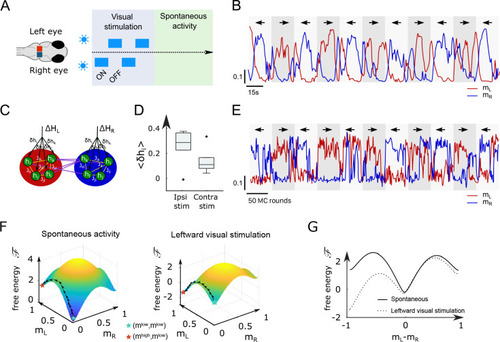
Modified Ising model captures the behavior of anterior rhombencephalic turning region (ARTR) under visual stimulation.

(A) Scheme of the stimulation protocol. The left and right eyes are stimulated alternatively for periods of 15–30 s, after which a period of spontaneous (no stimulus) activity is acquired. (B) Example of the ARTR activity signals under alternated left–right visual stimulation. The small arrows indicate the direction of the stimulus. (C) Sketch of the modified Ising model, with additional biases π›Ώβ„Žπ‘– to account for the local visual inputs. (D) Values of the additional biases averaged over the ipsilateral and contralateral (with respect to the illuminated eye) neural populations. n = 6 fish. (E) Monte Carlo activity traces generated with the modified Ising model. (F) Free-energy landscapes computed with the mean-field theory during spontaneous (left panel) and stimulated (right panel) activity for an example fish. (G) Free-energy along the optimal path as a function of π‘šπΏβˆ’π‘šπ‘… during spontaneous (plain line) and stimulated (dotted line) activity. The model is the same as in panel (F).

Expression Data

Expression Detail
Antibody Labeling
Phenotype Data

Phenotype Detail
Acknowledgments
This image is the copyrighted work of the attributed author or publisher, and ZFIN has permission only to display this image to its users. Additional permissions should be obtained from the applicable author or publisher of the image. Full text @ Elife