
Fig. 7
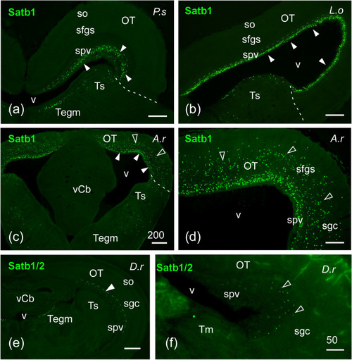
Photomicrographs of transverse single-labeled sections through mesencephalic areas of Polypterus senegalus (P.s), Lepisosteus oculatus (L.o), Acipenser ruthenus (A.r), and Danio rerio (D.r) showing the location of SATB1/2 or SATB1 immunoreactivity. The markers and the species are indicated in each image. a–f SATB1-ir cells in the periventricular layer of the optic tectum (spv), torus semicircularis and tegmentum of P. senegalus (a), L. oculatus (b), and A. ruthenus (c, d), the latter also with labeled cells in more superficial layers of the optic tectum; white arrowheads point to cells in spv, empty arrowheads in (c) and (d) point to cells in other tectal layers. e, f SATB1/2-ir cells in the optic tectum and tegmentum of D. rerio; arrowheads point to periventricular cells. For abbreviations, see list. Scale bars = 200 µm (c), 100 µm (a, b, d, e), 50 µm (f) |
| Antibody: | |
|---|---|
| Fish: | |
| Anatomical Terms: | |
| Stage: | Adult |