
FIGURE 1
- ID
- ZDB-FIG-230420-72
- Publication
- Santos-Ledo et al., 2022 - Oligodendrocyte origin and development in the zebrafish visual system
- Other Figures
- All Figure Page
- Back to All Figure Page
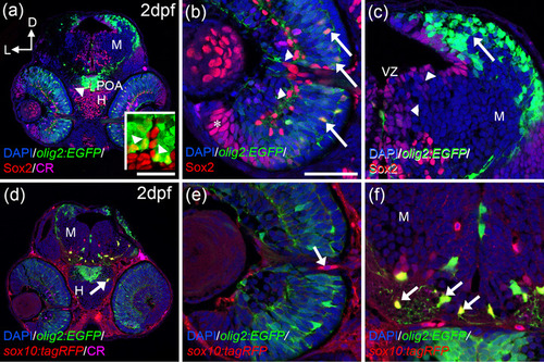
OPCs markers are detected from 2 dpf onwards. Distribution of olig2:EGFP/Sox2 (a–c) and olig2:EGFP/sox10:tagRFP (d–f) cells. olig2:EGFP and Sox2colocalize in the POA (arrowheads in a and inset in a), central outer retina (arrows in b), but not in the inner (arrowheads in c) or peripheral retina (asterisk in c). Olig2:EGFP cells are located in the dorsal optic tectum (arrow in c) but do not colocalize with Sox2 that is present in the VZ (arrowhead in c). sox10:tagRFP is absent from the retina except cells around the ON (d, arrow in e). In the hypothalamus and optic tectum, arborized olig2:EGFP cells also present sox10:tagRFP (arrows in d and f). Calretinin (CR) is present in several neurons, such as ganglion cells and is used to label the ON. D: dorsal; H: hypothalamus; L: lateral; M: mesencephalon; ON: optic nerve; POA: preoptic area; VZ: ventricular zone. Scale bar in a, d: 100 μm; in b, c, e, f: 50 μm |