
Fig. 9
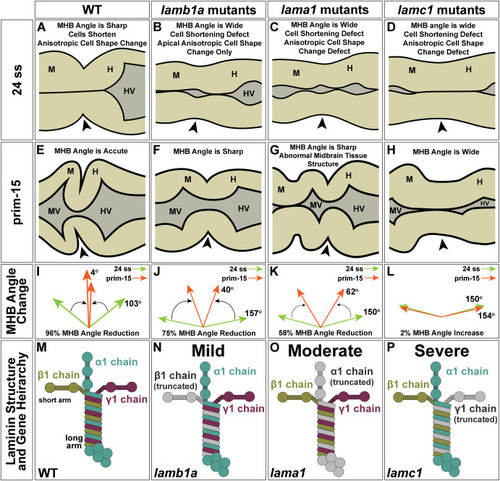
Laminin-111 mutant studies reveal a hierarchy within laminin-111 genes in their requirement for MHB basal tissue folding. (A–D) Schematic representation of MHB structure at 24 ss in WT (A) and lamb1a (B), lama1 (C), and lamc1 (D) mutants. Cell shape defects are indicated for each mutant. (E–H) Schematic representation of MHB structure at prim-15 in WT (E) and lamb1a (F), lama1 (G), and lamc1 (H) mutants. MHB angle defects are indicated for each mutant. (I–L) Diagram of changes in MHB angle from 24 ss to prim-15 in WT (I) and lamb1a (J), lama1 (K), and lamc1 (L) mutants. (M–P) Diagram of predicted laminin-111 protein structure and gene hierarchy between the laminin-111 genes. Arrowheads indicate MHBC. M; Midbrain; H, Hindbrain; MV, Midbrain Ventricle; HV, Hindbrain Ventricle. |
Reprinted from Developmental Biology, 492, Falat, E.J., Voit, G.C., Gutzman, J.H., Laminin-111 mutant studies reveal a hierarchy within laminin-111 genes in their requirement for basal epithelial tissue folding, 172-186, Copyright (2022) with permission from Elsevier. Full text @ Dev. Biol.