
Fig. 5
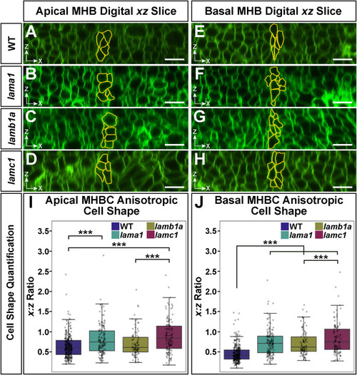
Individual loss of laminin-111 genes reveals a hierarchy in their requirement for apical and basal anisotropic cell shape change at the MHB. (A–H) Representative digital slice images of cells in the anterior-posterior (x-axis) and dorsal-ventral (z-axis) dimensions of embryos injected with memGFP and imaged at 24 ss. Cells at the MHBC are outlined in yellow within the apical region (A–D) and basal edge (E–H). (I–J) Apical and basal quantification of MHBC anisotropic cell shape (x:z ratio). Boxplots indicate the 25th and 75th percentiles and the median. At least three independent experiments are represented. n = 9 embryos for WT, 9 embryos for lama1 mutants, 6 embryos for lamb1a mutants, and 7 for lamc1 mutants. Significance values ∗∗∗P < 0.005. Scale bars: 20 μm. |
Reprinted from Developmental Biology, 492, Falat, E.J., Voit, G.C., Gutzman, J.H., Laminin-111 mutant studies reveal a hierarchy within laminin-111 genes in their requirement for basal epithelial tissue folding, 172-186, Copyright (2022) with permission from Elsevier. Full text @ Dev. Biol.