
Figure 1
- ID
- ZDB-FIG-230331-82
- Publication
- Watchon et al., 2023 - Autophagy Function and Benefits of Autophagy Induction in Models of Spinocerebellar Ataxia Type 3
- Other Figures
- All Figure Page
- Back to All Figure Page
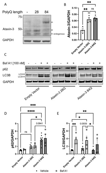
Autophagy impairment identified in a cell culture model of SCA3. (A) Western blot of SH-SY5Y cells stably expressing either an empty vector control or human ataxin-3 28Q or human ataxin-3 84Q probed for ataxin-3. (B) Quantification of human ataxin-3 levels relative to GAPDH showing increased levels of ataxin-3 in the ataxin-3 28Q and ataxin-3 84Q compared to the empty vector control (p = 0.0039 and p = 0.0019, respectively, n = 3–4). (C) Western blot of SH-SY5Y cells stably expressing either an empty vector control or human ataxin-3 28Q or human ataxin-3 84Q treated with either vehicle (DMSO) or Bafilomycin A1 (Baf A1). Western blot was probed with either p62 or LC3B. (D) Baseline levels of p62 were increased in ataxin-3 84Q cells compared to the empty vector control and ataxin-3 28Q cells (p < 0.0001 and p = 0.0301 respectively, n = 3–8). Baf A1 treatment increased p62 levels in the ataxin-3 84Q cells compared to the empty vector control (p = 0.0057).©) LC3II levels did not differ between genotypes at baseline; however, in the presence of Baf A1, empty vector cells had increased LC3II compared to vehicle treated empty vector cells (p = 0.0095). Vehicle-treated empty vector cells also had decreased LC3II levels compared to Baf A1-treated ataxin-3 28Q cells (p = 0.0193). Additionally, LC3II levels were lower in the ataxin-3 84Q cells treated with vehicle compared to the empty vector and ataxin-3 28Q in the presence of Baf A1 (p = 0.0069 and 0.0142 respectively, n = 4–8). Error bars represent mean ± SEM. Statistical analysis performed were a one-way ANOVA and two-way ANOVA followed by Tukey post hoc analysis. * represents p < 0.05, ** represents p < 0.01, *** represents p < 0.001, **** represents p < 0.0001. |