
Figure 6.
- ID
- ZDB-FIG-230321-7
- Publication
- Canham et al., 2023 - EVA1A (Eva-1 Homolog A) Promotes Endothelial Apoptosis and Inflammatory Activation Under Disturbed Flow Via Regulation of Autophagy
- Other Figures
- All Figure Page
- Back to All Figure Page
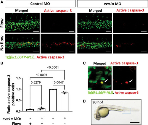
EVA1A (eva-1 homolog A) promotes endothelial apoptosis in vivo in zebrafish. A, Zebrafish embryos (transgenic flk1:EGFP-NLS embryos; green endothelial cell [EC] nuclei) were injected with morpholino oligonucleotide (MO) targeting eva1a or a nontargeting control MO. EC apoptosis was studied in the presence (control MO, flow) or in the absence (silent heart [sih] MO, no flow) of flow by whole-mount active caspase 3 staining (red). Apoptotic ECs (yellow) were monitored at 30 h postfertilisation (hpf). Lateral view, anterior to the left, dorsal up. B, The proportion of apoptotic ECs (number of apoptotic ECs divided by the total number of ECs) normalized to sih MO-injected embryos was calculated, and mean values are shown with SEM. Each data point represents an independent experiment with n≥5 embryos. Differences between groups were analyzed using a 2-way ANOVA with Tukey post hoc test and P values are shown in the graph. C, A high magnification image of EC apoptosis in a sih MO-injected embryo; white arrow indicates an apoptotic EC (yellow). D, Zebrafish embryo at 30 hpf. The region outlined with blue box represents the region that is studied in A. Scale bars: (A), 50 µm; (C), 10 µm; (D), 500 µm. |