
Fig. 7
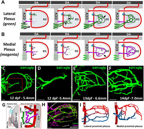
Formation of the proximal vascular plexus. (A,B) Schematics illustrating the growth and development of the lateral (A; in green) and medial (B; in magenta) proximal plexuses in association with the pectoral fin endoskeletal disk, occurring approximately between 6 mm and 8 mm larval length. (C-F) Confocal images of the pectoral fin vasculature of three separate Tg(kdrl:egfp)s843 larvae at 12 dpf (C,D), 13 dpf, (E) and 14 dpf (F). The red box in C indicates the approximate location of the higher-magnification images in D-F, which show the proximal plexus in progressively older animals. (G) Schematic illustrating the positioning of the lateral and medial pectoral proximal plexuses, (LPPP and MPPP, respectively) on either side of the endoskeletal disk within the larval pectoral fin. (H) Confocal image of an 18 dpf Tg(kdrl:egfp)s843 fish with the medial and lateral proximal plexuses false-colored in magenta and green, respectively. Note the vessels in the lateral plexus that disappear rostrally from the image. See Movie 13 for D image reconstructions of the same confocal stack shown in H. (I) Schematics illustrating the morphology and circulatory flow patterns of the lateral (left) and medial (right) proximal vascular plexuses shown in the confocal image in H. Red and blue labeling facilitates visualization of vessels supplying or draining the plexuses. Images shown are representative of data collected from ten separate larvae. DA, dorsal aorta; ED, endoskeletal disk; PA, pectoral artery; Pec fin, pectoral fin. Scale bars: 50 μm. |