Fig. 3
|
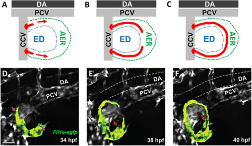
Formation of the primitive pectoral artery. (A-C) Schematics illustrating the stages of primitive pectoral artery formation, including initial dorsal and ventral sprouts from the CCV (A), elongation of these sprouts along the rim of the growing fin (B), and linkage of the two segments to form a complete primitive pectoral artery vascular arc (C). DA, dorsal aorta; ED, endoskeletal disk; PCV, posterior cardinal vein. (D-F) Still images taken from a time-lapse confocal time series of primitive pectoral artery formation in a Tg(fli1a:egfp)y1 transgenic zebrafish at 34 hpf (D), 38 hpf (E) and 40 hpf (F). Pink arrows indicate the emerging sprouts migrating into the pectoral fin, and red arrows indicate the emerging ventral arterial sprout. See Movie 2 for the complete time-lapse series. Images shown are representative of data collected from 12 separate embryos. Scale bar: 40 μm (D-F). |

