
Fig 6
- ID
- ZDB-FIG-230315-56
- Publication
- Xie et al., 2023 - Ependymal polarity defects coupled with disorganized ciliary beating drive abnormal cerebrospinal fluid flow and spine curvature in zebrafish
- Other Figures
- All Figure Page
- Back to All Figure Page
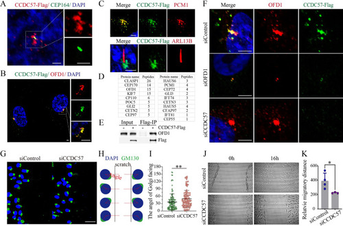
CCDC57 is a centrosomal satellite protein required for cell polarity.
(A-C) Confocal images showing the relative localization of CCDC57 with OFD1, PCM1, CEP164, and ARL13B in RPE-1 cells. (D) Mass spectrometry analysis of the CCDC57 interacting proteins. (E) Immunoprecipitation results showing the interaction between CCDC57 and OFD1. (F) Confocal images showing the localization of CCDC57 and OFD1 in siRNA knockdown RPE-1 cells. (G) Scratch-wound assay showing the polarized localization of Golgi (GM130, green) during directional cell migration in control and CCDC57 siRNA-treated cells. (H) Model illustrating the statistical analysis of cell polarity by Golgi position. (I) Dot plots showing the angles of Golgi facing the migration edge in control and CCDC57 siRNA-treated cells. (J, K) Images showing the cell migration state and statistical analysis of the migration distance. In all panels, nuclei were counterstained with DAPI in blue. Scale bars: 2.5 μm in panel A; 5 μm in panel B; 3 μm in panel C; 5 μm in panel F; 50 μm in panel G. The data underlying the graphs shown in the figure can be found in S1 Data. |