Fig. 4
- ID
- ZDB-FIG-230312-32
- Publication
- Nam et al., 2022 - The Novel Human HtrA2 Ortholog in Zebrafish: New Molecular Insight and Challenges into the Imbalance of Homeostasis
- Other Figures
- All Figure Page
- Back to All Figure Page
|
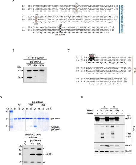
Fig. 4. Enzymatic conservation of zHtrA2 as a serine protease. (A) The sequence alignment for identifying the putative serine protease domain (AA 143–319) of zHtrA2. The catalytic triad residues are indicated by boxes. The corresponding domain of hHtrA2, i.e. residues 166–342, was used as an alignment control. Identical residues (“*”), residues with highly similar properties of the R group (“:”), and residues with weakly similar properties of the side chain (“.”) were denoted. (B) Autocatalytic processing of pro-zHtrA2. The indicated pro-zHtrA2 proteins, which were synthesized using an in vitro TNT SP6 system, were probed with D-Tag Ab. (C) Sequence alignment of the putative PDZ domain (AA 337–423) of zHtrA2. The XΦGΦ motif in the PDZ plays a key role in ligand binding, where G is highly conserved, and X and Φ represent any and hydrophobic AAs, respectively. (D) Endoproteolytic activity of zHtrA2. The level of zHtrA2 bound to the anti-FLAG beads was probed with D-Tag Ab. The immunoglobulin heavy (IgH; “*”) and light (IgL; “**”) chains are indicated. (E) Proteolytic cleavage of the hHtrA2’s cognate substrate, Parkin, by zHtrA2. The cleavage pattern of Parkin (17 and 19 kDa) was analyzed with the Parkin Ab. zHtrA2 was detected by D-Tag Ab. |
Reprinted from Gene, 819, Nam, M.K., Moon, J.M., Kim, G.Y., Min Kim, S., Rhim, H., The Novel Human HtrA2 Ortholog in Zebrafish: New Molecular Insight and Challenges into the Imbalance of Homeostasis, 146263, Copyright (2022) with permission from Elsevier. Full text @ Gene

