
Figure 5
- ID
- ZDB-FIG-230228-225
- Publication
- Prykhozhij et al., 2023 - Loss of calpain3b in Zebrafish, a Model of Limb-Girdle Muscular Dystrophy, Increases Susceptibility to Muscle Defects Due to Elevated Muscle Activity
- Other Figures
- All Figure Page
- Back to All Figure Page
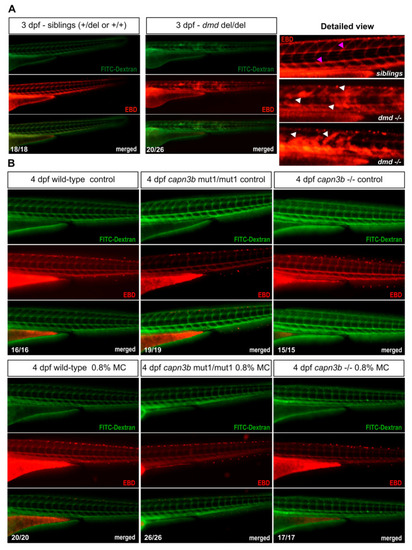
Evans Blue Dye analysis shows lack of permeability disruption by methylcellulose incubation in both wild-type and capn3b mutant embryos. (A) Evans Blue Dye (EBD) and FITC-Dextran injected sibling and dmd−/− embryos at 3 dpf. EBD signal is labeled in red and FITC-dextran in green. Merged images suggest a near complete co-localization of EBD and FITC-dextran. Vascular labeling indicates the normal situation where muscle permeability is not disrupted, whereas the more widespread labeling indicates disruption of muscle cell permeability. The detailed magnified view shows examples of sibling and dmd−/− embryo EBD labeling. White arrowheads show damaged muscle labeling and fuchsia arrowheads label EBD in the blood vessels in the sibling embryos. (B) EBD assay of wild-type, capn3bmut1/mut1, and capn3b−/− mutant embryos after culturing them from 2 days post-fertilization (dpf) to 4 dpf under control conditions or in 0.8% methylcellulose (MC) in E3 medium. Only vascular labeling could be detected in all of the imaged embryos. The combined numbers out of the total analyzed are shown in the bottom right-hand corner of the representative images. |
| Fish: | |
|---|---|
| Condition: | |
| Observed In: | |
| Stage Range: | Protruding-mouth to Day 4 |