
FIGURE 7
- ID
- ZDB-FIG-230228-112
- Publication
- Boyd et al., 2023 - Clcf1/Crlf1a-mediated signaling is neuroprotective and required for Müller glia proliferation in the light-damaged zebrafish retina
- Other Figures
- All Figure Page
- Back to All Figure Page
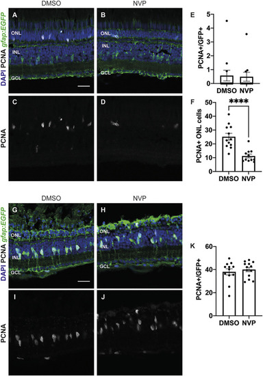
CLCF/CRLF1 induces rod precursor cell proliferation in an IGF-1R dependent manner. (A–D) Single confocal images from albino; Tg[gfap:EGFP] nt11 zebrafish which were intravitreally injected every 12 h with CLCF1/CRLF1 and simultaneously receiving injections of either DMSO (vehicle) (A) or the IGF-1R inhibitor NVP (B) for 72 h. Sections were labeled for GFP and PCNA to assess proliferating Müller glia and nuclei were counterstained with DAPI (C, D). (E) Quantification showing no significant change in the number of PCNA-positive Müller glia within the INL of NVP-treated fish. Student’s t-test, p = 0.87, n ≥ 11. (F) Quantification showing significant decrease in the number of PCNA-positive cells within the ONL of NVP-treated fish. Student’s t-test, p = 0.0001, n ≥ 11. (G–J) Single confocal images from dark-adapted albino; Tg[gfap:EGFP] nt11 zebrafish which were light-treated for 36 h while receiving injections of either DMSO (vehicle) (G) or the IGF-1R inhibitor NVP (H). Sections were labeled for GFP and PCNA to assess proliferating Müller glia and nuclei were counterstained with DAPI (I, J). (K) Quantification showing no significant change in the number of PCNA-positive Müller glia between DMSO and NVP-treated fish. Student’s t-test, p = 0.54, n ≥ 12. Mean ± SEM, ****p < 0.0001. ONL, outer nuclear layer, INL, inner nuclear layer, GCL, ganglion cell layer. Scale bar represents 20 µm. |