
Fig. 2
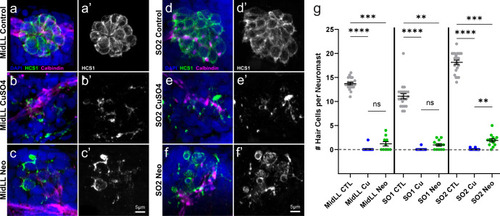
Confirmation of neuromast hair cell loss following CuSO4 or neomycin treatment.
a–f Representative confocal max intensity projection images of the: a–c) mid posterior lateral line (MidLL) fourth neuromast (L4); and d–f second anterior supraorbital (SO2) neuromast from the fish cohorts used for behavior experiments. Hair cells were labeled with an antibody against Otoferlin (HCS1; green a–f, gray a′–f′). Afferent neurons were labeled with an antibody against Calbindin (magenta), and cell nuclei were labeled with DAPI (blue). g Quantification of the grand mean (±SEM) number of hair cells per neuromast in intact (CTL), CuSO4- and neomycin-treated fish. Each dot represents the mean number of hair cells from the MidLL (L3, L4, and L5) or SO (left and right) neuromasts from an individual fish. Data were collected from fish used in three experimental behavior trials; n = 4–6 fish per condition per trial. Significance values: **<0.01, ***<0.001, ****<0.0001. |