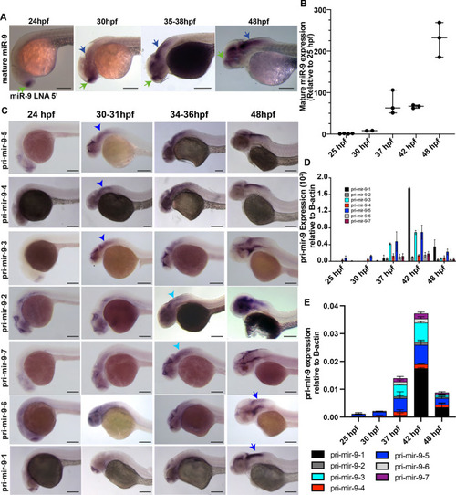
Pri-mir-9 paralogues are expressed with different temporal onset. (A) Representative example of chromogenic whole-mount in situ hybridization (WM-ISH) of miR-9 using miR-9 LNA 5′-Dig observed at different stages during development. Similar results can be observed in Soto et al. (2020). Longitudinal view, anterior to the left. Green arrow, forebrain expression; blue arrow, hindbrain expression. (B) Taqman RT-qPCR of mature miR-9 from dissected hindbrain at different stages of development, relative to 25 hpf. Horizontal bars indicate median with 95% confidence intervals. (C) Chromogenic WM-ISH of different pri-mir-9s using specific probes for each paralogue observed at different stages during development. Longitudinal view, anterior to the left. Blue arrowhead, expression in hindbrain at 30-31 hpf; light blue arrowhead, expression in hindbrain at 34-36 hpf; blue arrow, expression in hindbrain at 48 hpf. (D,E) SYBR green RT-qPCR relative quantification of the seven pri-mir-9s from dissected hindbrains at different stages of development. Quantification was normalised using β-actin. Data are mean±s.d. (B,D,E) N=3, each N contains a pool of 10 hindbrains. Scale bars: 200 μm.
|
