
Figure 3
- ID
- ZDB-FIG-221230-12
- Publication
- Stenzel et al., 2022 - Distinct and redundant roles for zebrafish her genes during mineralization and craniofacial patterning
- Other Figures
- All Figure Page
- Back to All Figure Page
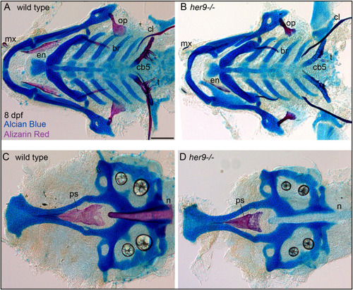
Mineralization defects in her9 homozygous mutants partially recover at late stages of larval development. Zebrafish heterozygous for her9 were pairwise intercrossed and at eight days post fertilization (dpf) larvae were stained with Alcian Blue and Alizarin Red to label cartilage and bone. The individuals were then genotyped, the viscerocrania and neurocrania were dissected, flat mounted, and then imaged. (A, B) Viscerocrania from wild-type and her9 homozygous mutant larvae. (C, D) Neurocrania from wild-type and her9 homozygous mutant larvae. The following craniofacial elements are indicated: opercle bone (op), branchiostegal ray (br), maxilla (mx), entopterygoid (en), ceratobranchial 5 (cb5) bone, teeth (t), cleithrum (cl), parasphenoid bone (ps), and notochord (n). Scale bar is 100 μm. |
| Fish: | |
|---|---|
| Observed In: | |
| Stage: | Days 7-13 |