Figure 8
- ID
- ZDB-FIG-221226-170
- Publication
- López-Cuevas et al., 2022 - Macrophage Reprogramming with Anti-miR223-Loaded Artificial Protocells Enhances In Vivo Cancer Therapeutic Potential
- Other Figures
- All Figure Page
- Back to All Figure Page
|
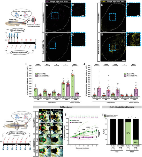
Anti‐miR223 protocell treatment reduces cancer progression in adult zebrafish. A–H) Single‐channel confocal images of immunostained cryosections from 1‐year‐old adult tail tumors at 3 dpt after a single local injection of unlabeled control protocells (A,B,E,F) or unlabeled anti‐miR223 protocells (C,D,G,H); white lines indicate tumor margins; proliferating cells are revealed by anti‐pH3 immunostaining (magenta) in (A)–(D) and apoptotic cells revealed by TUNEL staining (yellow) in (E)–(H). B,D,F,H) High magnification views of (A), (C), (E), and (G). I,J) Graphs showing percentage of pH3‐positive (I) or TUNEL‐positive (J) area over total tumor area from sections after each injection regime quantified from the regions imaged in (A), (C), (E), and (G). K–R) Multi‐channel (K,M,O,Q) or single‐channel (L,N,P,R) images of 1‐year‐old adult tail tumors (red outlines) at 0 and 30 dpt after multiple local injections of unlabeled control protocells (K–N) or unlabeled anti‐miR223 protocells (O–R). S) Graph showing adult tail tumor growth curves after each protocell treatment quantified from the regions imaged in (K)‐(R) and (1). See also Figure |

