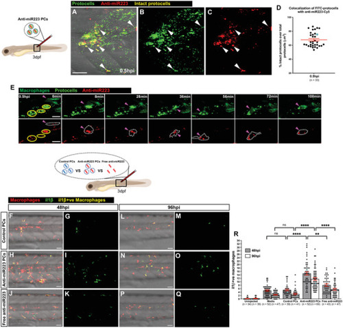
Uptake of anti‐miR223 protocells enhances il1β expression in macrophages. A–C) Multi‐channel (A) or single‐channel (B,C) confocal images of the flank of a 3 dpf casper larva after local injection of anti‐miR223‐Cy5 FITC‐protocells at 0.5 hpi; white arrowheads indicate anti‐miR223 protocells that remain intact post injection. D) Dot plot showing percentage of intact protocells as quantified by colocalization of FITC (protocells) with Cy5 (anti‐miR223) from the regions imaged in (A)–(C). E) Single‐channel confocal movie frames of a region of the flank of a 3 dpf Tg(mpeg1:FRET) larva after local injection of anti‐miR223‐Cy5 FITC‐protocells at 0.5 hpi showing the uptake of intact protocells (yellow circles) by a macrophage (magenta arrowheads and white dotted outlines). See also Movie S10, Supporting Information. F–Q) Multi‐channel (F,H,J,L,N,P) or single‐channel (G,I,K,M,O,Q) confocal images of the flank of Tg(mpeg1:mCherry;il1β:GFP) larvae showing il1β‐positive macrophages (yellow) (il1β‐negative macrophages are red) after local injection of unlabeled control protocells, unlabeled anti‐miR223 protocells or unlabeled free anti‐miR223 at 3 dpf and imaged at 48 hpi (F–K) and 96 hpi (L–Q). R) Graph showing the number of il1β‐positive macrophages following each treatment quantified from the regions imaged in (F)–(Q). See also Figures S7 and S8, Supporting Information. Accompanying schematics illustrate developmental stage (larva), type of injection (local), and imaged area (black outlined box) used for each experiment. Data are pooled from three independent experiments and analyzed using Kruskal–Wallis test with Dunn's multiple comparisons test (R), ns p ≥ 0.05, **p < 0.01, ****p < 0.0001. Graphs show mean ± SEM, each dot represents one fish and blue dots correspond to the representative images shown in the panels. n = number of fish; PCs = protocells. Scale bars = 50 µm (A,F,H,J,L,N,P), 20 µm (E).
|

