
FIGURE
Fig. 4
- ID
- ZDB-FIG-221216-14
- Publication
- Spelat et al., 2022 - Metabolic reprogramming and membrane glycan remodeling as potential drivers of zebrafish heart regeneration
- Other Figures
- All Figure Page
- Back to All Figure Page
Fig. 4
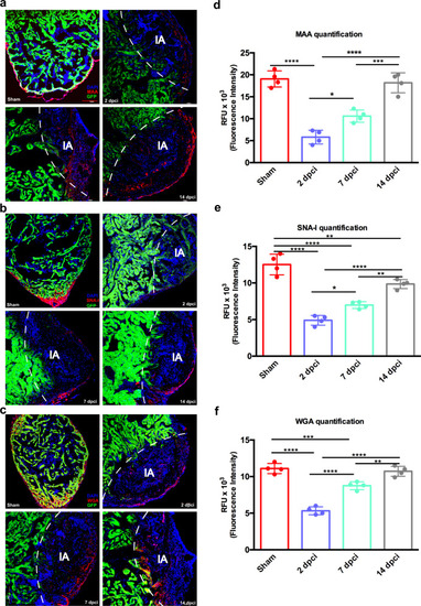
Sialic acid distribution in regenerating zebrafish heart identified by lectin histochemistry.
a Lectin histochemistry showing the distribution of MAA, recognizing α-(2,3)-linked sialic acid, b SNA-I, binding to α-(2,6)-linked sialic acid and c WGA, binding to sialic acid and GlcNAc, in ventricle tissue. d MAA, e SNA-I and f WGA relative quantification. The lectin binding quantifications highlighted a similar decrease at 2 dpci and a subsequent increase in the following timepoints. Cardiomyocytes were identified by GFP positivity. IA injured area. Data are shown as mean with SD (n = 4 animals per group). Scale bar = 100 μm. *p < 0.05, **p < 0.01, ***p < 0.001, ****p < 0.0001. |
Expression Data
Expression Detail
Antibody Labeling
Phenotype Data
Phenotype Detail
Acknowledgments
This image is the copyrighted work of the attributed author or publisher, and
ZFIN has permission only to display this image to its users.
Additional permissions should be obtained from the applicable author or publisher of the image.
Full text @ Commun Biol