FIGURE

Figure 5

ID
ZDB-FIG-221214-214
Publication
Lee et al., 2022 - Evaluation of Cisplatin-Induced Pathology in the Larval Zebrafish Lateral Line
Other Figures
All Figure Page
Back to All Figure Page
Figure 5

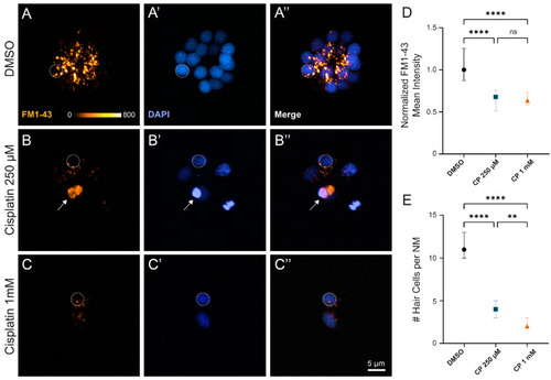
Surviving neuromast hair cells after cisplatin treatment demonstrate impaired mechanotransduction. (AC) Maximum-intensity projections of confocal images show wildtype zebrafish neuromasts after 2 h treatment with 0.1% DMSO (A), 250 µM cisplatin (B), and 1 mM cisplatin (C) followed by a 24 h recovery in EM. Dotted circles exemplify 4 µm region of interest used to measure mean FM1-43 intensity. Arrow indicates non-viable hair cell that was not included in analysis (B). Mean hair cell FM1-43 intensity (normalized to control) demonstrates decreased mechanotransduction among cisplatin-treated fish compared to those exposed to vehicle control ((D), **** p < 0.0001, Kruskal–Wallis test). Post hoc analysis with Dunnett’s multiple comparisons test failed to detect significant changes in FM1-43 uptake between moderate and high dose cisplatin ((D), ns > 0.9999). Cisplatin demonstrates a dose-dependent increase in hair cell loss ((E), **** p < 0.0001, ** p = 0.0069; Dunnett’s multiple comparisons test). Median values with corresponding 95% confidence intervals shown for both FM1-43 uptake and hair cell counts. n = 3–4 fish (2–3 neuromasts per fish). N = 3 trials.

Expression Data

Expression Detail
Antibody Labeling
Phenotype Data

Phenotype Detail
Acknowledgments
This image is the copyrighted work of the attributed author or publisher, and ZFIN has permission only to display this image to its users. Additional permissions should be obtained from the applicable author or publisher of the image. Full text @ Int. J. Mol. Sci.