
Fig. 7
- ID
- ZDB-FIG-221211-73
- Publication
- Ablain et al., 2022 - Loss of NECTIN1 triggers melanoma dissemination upon local IGF1 depletion
- Other Figures
- All Figure Page
- Back to All Figure Page
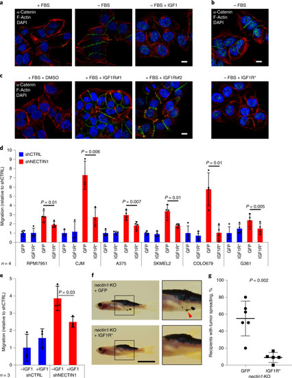
a, Immunofluorescence analysis of α-catenin (green) and F-actin (red) in A375 human melanoma cells in the presence or absence of serum (FBS) (blue, DAPI), complemented or not with 100 ng ml−1 IGF1 for 12 h. Scale bar, 10 μm. Data are representative of four independent experiments. b, Immunofluorescence analysis of α-catenin (green) and F-actin (red) in A375 human melanoma cells expressing or not a constitutively active form of the IGF1 receptor (CD8-IGF1R, IGF1R*), and cultured in the absence of serum (FBS) for 12 h (blue, DAPI). Scale bar, 10 μm. Data are representative of three independent experiments. c, Immunofluorescence analysis of α-catenin (green) and F-actin (red) in A375 human melanoma cells cultured in the presence of serum (FBS) and treated with DMSO (vehicle) or with two different IGF1R inhibitors (IGF1Ri#1: Linsitinib, 1 μM; IGF1Ri#2: GSK1838705A, 1 μM) for 12 h (blue, DAPI). Scale bar, 10 μm. Data are representative of four independent experiments. d, Migration of six human melanoma cell lines conditionally expressing GFP or a constitutively active form of IGF1R (CD8-IGF1R, IGF1R*), and stably expressing an shRNA directed against NECTIN1 (shNECTIN1) relative to cells expressing a control shRNA (shCTRL) in a transwell assay. Cells were allowed to migrate for different times depending on the cell line (Extended Data Fig. 4i and Methods). Data represent mean ± s.d. of four independent experiments (paired two-tailed t-test). e, Migration of A375 human melanoma cells stably expressing either a control shRNA (shCTRL) or an shRNA directed against NECTIN1 (shNECTIN1) in a transwell assay after 12 h of serum starvation in the presence or absence of 100 ng ml−1 IGF1. Cells were allowed to migrate for 6 h. Data represent mean ± s.d. of three independent experiments (paired two-tailed t-test). f, Representative images of adult casper zebrafish transplanted with nectin1-knockout primary zebrafish melanoma cells expressing GFP or a constitutively active form of IGF1R (CD8-IGF1R, IGF1R*), 21 days after injection. Insets show ×2 magnification views. Arrowhead indicates patches of disseminated melanoma cells. Scale bar, 1 cm. g, Quantification of the proportion of secondary recipients of nectin1-knockout tumors expressing GFP (n = 6) or nectin1-knockout tumors expressing CD8-IGF1R (IGF1R*) (n = 5) showing tumor spreading. Data represent mean ± s.d. (two-tailed t-test). |