
Figure 2
- ID
- ZDB-FIG-221118-139
- Publication
- Greco et al., 2022 - PCSK9 Confers Inflammatory Properties to Extracellular Vesicles Released by Vascular Smooth Muscle Cells
- Other Figures
- All Figure Page
- Back to All Figure Page
|
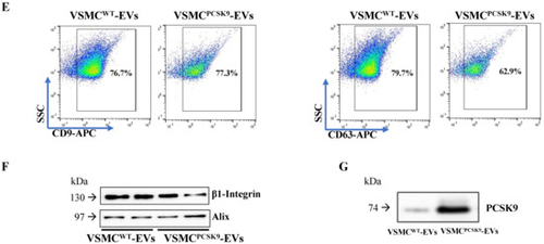
Phenotypic characterization of extracellular vesicles (EVs). (A) Cell viability of VSMCs supplemented or not with FBS was assessed by annexin V/propidium iodide double staining. n = 9 per group. (B) EV concentrations are expressed as means for each size for the VSMCPCSK9-EVs (dashed line) and VSMCWT-EVs (solid line). EV concentrations were calculated as marginal means from negative binomial regression models. n = 6 derived from 6 different experiments. (C) p-value and False Discovery Rate p-value of the comparisons of each size EV for the entire 30–700 nm size range are reported. n = 6 derived from 6 different experiments. (D) Representative ultrastructural images of EVs by TEM. For both, the main image shows an EV whose size is compatible with intermediate EVs, and the insert shows an EV whose size is compatible with small EVs (arrow). Scale bar = 100 nm. (E) The expression of tetraspanins CD9 and CD63 was evaluated by flow cytometry analysis in EVs. Representative panels of three independent experiments. (F) Protein expression of β1-integrin and Alix as assessed by WB in EVs. Representative blots of three independent experiments. (G) PCSK9 expression in VSMCWT-EVs and VSMCPCSK9-EVs through WB analysis. Representative blot of three independent experiments. VSMCPCSK9-EVs are those released by VSMCPCSK9 and VSMCWT-EVs are those released by VSMCsWT. EV, extracellular vesicles; FBS, foetal bovine serum; VSMCPCSK9-EVs, EVs released by VSMCs overexpressing PCSK9; VSMCWT-EVs, EVs released by VSMCs wildtype; TEM, Transmission electron microscopy; WB, Western blot. 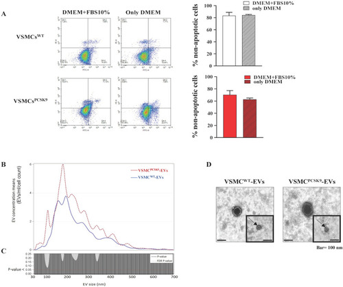
|