Figure 1
- ID
- ZDB-FIG-221031-48
- Publication
- Jiang et al., 2022 - Cloning, Exogenous Expression and Function Analysis of Interferon-γ from Gadus macrocephalus
- Other Figures
- All Figure Page
- Back to All Figure Page
|
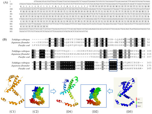
Bioinformatic analysis of GmIFN–γ gene. (A) cDNA and the deduced amino acid of GmIFN–γ. Signal peptide was underlined. (B) Mature IFN–γ peptides among Takifugu rubripes, Japanese flounder and G. macrocephalus were aligned. Identical or similar amino acids are shaded (black: present in all species; dark gray: present in 80% of the species). The nuclear localization signal and the putative IFN–γ signature sequence are boxed in green. (C1–D3): 3D models of IFN–γ compared between Japanese flounder and G. macrocephalus. (C1,C2): a 3D model of Japanese flounder displayed in Ribbons and Spacefill style, separately. PDB code is c6f1eA. (D1,D2): a 3D model of GmIFN–γ was constructed based on the template c6f1eA. Both were displayed in Ribbons and Spacefill style, separately. (D3) Secondary structure assignment was carried using PROCHECK. Good quality is indicated in blue, while the bad quality is indicated in red. |

