
Figure 4
- ID
- ZDB-FIG-221029-25
- Publication
- Davison et al., 2022 - Zebrafish Slit2 and Slit3 Act Together to Regulate Retinal Axon Crossing at the Midline
- Other Figures
- All Figure Page
- Back to All Figure Page
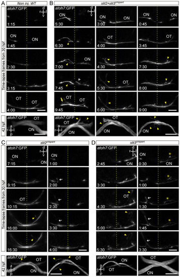
Retinal axons from slit2, slit3 and slit2/slit3 double crispants present navigation errors around the midline. Confocal time lapse observations of pioneer optic axon growth along the area surrounding the optic chiasm, in atoh7:Gap43-EGFP (atoh7:GFP) transgenic embryos. (A–D). Sequences of selected frames from time-lapse analysis of non-injected wild-type (A), slit2 + slit3 crispant (B), slit2 crispant (C) and slit3 crispant (D) embryos. A frontal view of the chiasm area is shown, starting with the appearance of the first axon at the optic nerve. The dashed lines indicate the location of the midline. Maximum intensity z-projections of the corresponding embryos are shown at the bottom of each time-lapse sequence, in ventral view. Misrouted axons were classified and quantified (Table 2) as follows: projections to the ipsilateral optic tract (e.g., white arrows), projections to the contralateral optic nerve (e.g., yellow arrow), fasciculation defects (e.g., asterisks), and immediately corrected minor pathfinding errors (e.g., empty white arrowhead). Yellow arrowheads point to other errors seen in these images. ON: optic nerve; OT: optic tract. Scale bars: 30 µm. See Videos S4–S8. |