|
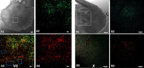
Bucky ball and CVH are no longer co-localized within the nuclear region of primordial germ cells during the area pellucida formation. 7A, in EGK VII embryos, at the beginning of area pellucida formation, Bucky ball (green) in the center of the germinal disc diffused back into the cytoplasm (A2), while CVH (red, A4) is contained predominantly in the nuclei. This is visualized clearly in the overlay (A3) as a red center of the germline predisposed cells surrounded by the green Bucky ball fluorescence. Blue frames with asterisk indicate only CVH labeled without Bucky ball at the lateral edges of the central region. DAPI as nuclear marker visualizes the central concentration of the germline cells and as white overlay the nuclear localization of CVH and DAPI in the germline cells. This pattern is stable until oviposition at stage X (B). Scales in (A1) = 200 µm, (B1): 500 µm, (A2–A4) 20 µm, (B2–B4): 100 µm.
|

