
FIGURE
Figure 7
Figure 7
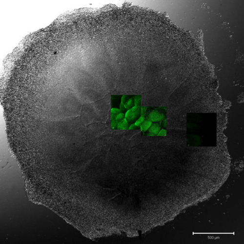
Overview of an intrauterine embryo (EGK III) with two frames of the central region with Bucky ball immunolabeling in contrast to severely reduced immuno-fluorescence at the lateral outline of furrows. The two frames in the central region show the cells with Bucky ball immunofluorescence in cytoplasmic granular distribution. The lateral frame, documented at the exactly comparable acquisition conditions, does not provide signals above background. Scale = 500 µm. |
Expression Data
Expression Detail
Antibody Labeling
Phenotype Data
Phenotype Detail
Acknowledgments
This image is the copyrighted work of the attributed author or publisher, and
ZFIN has permission only to display this image to its users.
Additional permissions should be obtained from the applicable author or publisher of the image.
Full text @ Sci. Rep.