
FIGURE 1
- ID
- ZDB-FIG-221018-143
- Publication
- Agostini et al., 2022 - Conserved and diverged asymmetric gene expression in the brain of teleosts
- Other Figures
- All Figure Page
- Back to All Figure Page
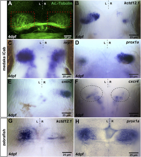
Visualisation of asymmetries in the medaka habenulae. iCab embryos at four dpf focused on the dorsal diencephalon; anterior to the top. (A) Immunostaining for anti-acetylated tubulin shows subtle left-right asymmetric dendritic arborisation in the habenula. Please compare the green signal within the same-sized red boxes. (B–H) Expression of habenula and pineal complex marker genes. (B,C) kctd12.1 and nrp1 are stronger expressed in left-sided habenular neurons. (D,E) prox1a and cntn2 are mainly expressed in right-sided habenular neurons, while (F) cxcr4b is expressed in habenular progenitors, the area of which is encircled. (G) In the zebrafish, kctd12.1 is expressed in lateral neurons of the dorsal habenulae, while (H) prox1a is expressed in medial dorsal habenular neurons. |