
FIGURE
Fig. 1
- ID
- ZDB-FIG-221018-1
- Publication
- Garcia-Concejo et al., 2021 - Protein kinase C family evolution in jawed vertebrates
- Other Figures
- All Figure Page
- Back to All Figure Page
Fig. 1
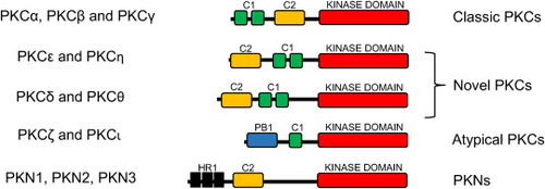
Fig. 1. Structure and classification of PKC subfamilies. The C1 domain, present in all subfamilies except for PKN, is formed by two zinc-finger motifs and is responsible for binding DAG and phorbol esters. The C2 domain (missing in the atypical PKCs) mediates calcium response. The PB1 domain, only present in the atypical PKCs, provides specificity during intracellular signaling. The HR1 domains, only found in the PKNs, mediate activation by Rho proteins. |
Expression Data
Expression Detail
Antibody Labeling
Phenotype Data
Phenotype Detail
Acknowledgments
This image is the copyrighted work of the attributed author or publisher, and
ZFIN has permission only to display this image to its users.
Additional permissions should be obtained from the applicable author or publisher of the image.
Reprinted from Developmental Biology, 479, Garcia-Concejo, A., Larhammar, D., Protein kinase C family evolution in jawed vertebrates, 77-90, Copyright (2021) with permission from Elsevier. Full text @ Dev. Biol.