
Fig. 4
- ID
- ZDB-FIG-220921-85
- Publication
- Dunn et al., 2022 - Comparative in situ hybridization protocols in zebrafish
- Other Figures
- All Figure Page
- Back to All Figure Page
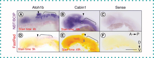
Figure 4. Single in situ hybridization for atoh1b demonstrates the superiority of NBT/BCIP as a stain. (A–C) NBT/BCIP is more sensitive and offers higher contrast. (D–F) Fast Red displays less nonspecific staining in red, although discolors the otherwise clear embryo yellow. (A & D)Atoh1b is expressed in the rhombic lip (filled arrowhead) and hindbrain (bracket) in zebrafish embryos at 24 h postfertilization. Both stains label the predicted region and display paler, nonspecific staining, particularly in the eye. (B & E)Cabin1 is expressed in many brain regions, including the ventral rhombic lip (open arrowheads) and hindbrain (bracket). (C & F) Sense probes for Cabin1 served as controls. Embryos were manually deyolked. Scale bar = 100 μm. |