
Fig. 2
- ID
- ZDB-FIG-220908-31
- Publication
- Chen et al., 2022 - Cysteamine affects skeletal development and impairs motor behavior in zebrafish
- Other Figures
- All Figure Page
- Back to All Figure Page
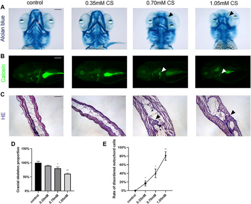
Skeletal staining of embryos after exposure to cysteamine. (A) Alcian blue staining of zebrafish larvae craniofacial skeleton exposed to 0.35, 0.70, and 1.05 mM cysteamine at 72 hpf. (B) Calcein staining of the whole-mount skeleton of zebrafish larvae exposed to 0.35, 0.70, and 1.05 mM cysteamine at 120 hpf. Arrows indicated abnormal craniofacial skeletal structure and chondropenia. mc: meckel’s cartilage, ch: ceratohyals. (C) HE staining of the skeleton of zebrafish larvae exposed to 0.35, 0.70, and 1.05 mM cysteamine at 72 hpf. Arrows indicated disordered accumulation of notochord cells and abnormal tissue vacuoles. (D) The craniofacial skeleton area of zebrafish larvae exposed to 0.35, 0.70, and 1.05 mM cysteamineat 72 hpf. (E) Rate of disordered notochord cells in zebrafish larvae exposed to 0.35, 0.70, and 1.05 mM cysteamine at 72 hpf. Scale bars: 50 μm (A,C), 100 μm (B). *p < 0.05, **p < 0.01, ***p < 0.001, mean ± S.D. |