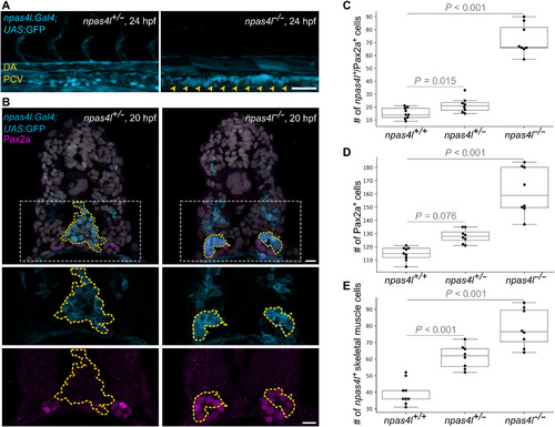
Fig. 2. npas4l reporter–expressing cells are observed in pronephron tubules and skeletal muscle in npas4l mutants. (A) npas4l reporter expression in ventral cells of 24 hpf npas4l+/− (left) and npas4l−/− (right) embryos. The main axial vessels (DA and PCV) are visible in npas4l+/− embryos, while only the more ventrally located cells are detected in npas4l−/− embryos (arrowheads). (B) Transverse sections of 20 hpf npas4l+/− (left) and npas4l−/− (right) embryos. The endothelial progenitors (outlined by yellow dotted lines) in npas4l mutants fail to reach the midline unlike those in wild-type siblings; they also remain round. The round ventrolateral npas4l reporter–expressing cells in npas4l−/− embryos also express Pax2a and are part of the pronephron tubules. (C to E) Numbers of npas4l reporter/Pax2a double-positive cells (C), all Pax2a-positive cells (D), and npas4l reporter–expressing skeletal muscle cells (E) per field of view (319.45-μm-long area over the yolk extension). Cells were counted in three-dimensional (3D) confocal lateral views of npas4l+/+, npas4l+/−, and npas4l−/− embryos at 24 hpf. Data are represented as individual data points, median, interquartile range, and extremes excluding outliers. P values were calculated by Poisson regression and adjusted to counteract the multiple testing problem. Data represent a subset of the data shown in Fig. 6 (B to D). DA, dorsal aorta; PCV, posterior cardinal vein. Scale bars, 50 μm (A) and 10 μm (B).
|
