|
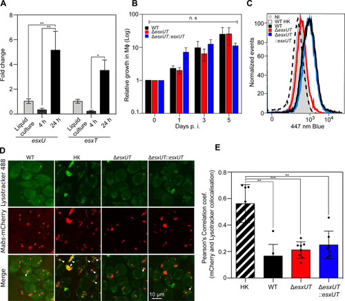
Δ<italic toggle='yes'>esxUT</italic> is maintained in an intact phagosome without acidification in macrophages.(A) Expression of esxU and esxT genes in J774.2 macrophages at 4 hrs and 24 hrs post-infection (black) relative to planktonic growth expression in broth medium (gray). (B) Relative intracellular survival of WT (black), ΔesxUT (red), and ΔesxUT::esxUT (blue) strains as determined by CFU counts during infection in J774.2 macrophages at an MOI of 10:1. (C) Phagosomal rupture detected by CCF-4 FRET-based flow cytometry 24 hrs post-infection. Results are depicted as signal overlays per group with 1,000,000 events per condition acquired in WT (black line), heat-killed (dotted line), ΔesxUT (red line), ΔesxUT::esxUT (blue line) strains and in uninfected cells (NI, gray line). (D) Colocalization of M. abscessus WT, heat-killed, ΔesxUT and ΔesxUT::esxUT strain expressing mCherry with the acidotropic dye LysoTracker Green in infected J774.2 cells 3 hrs post-infection. Arrows indicate intracellular mycobacteria co-localizing with LysoTracker green. (Scale bar, 10 μm). (E) mCherry-labeled strains colocalized with LysoTracker were measured by Pearson correlation of at least 100 infected cells in 10 different fields. Data are representative of three (B and C) or two (D and E) independent experiments and represent means ± SEM. P values were determined by ANOVA with Tukey’s test using GraphPad prism program (A and B) and unpaired t test (E); ns, not significant; *P < 0.05, **P < 0.01, ***P < 0.001, ****P < 0.0001.
|